Download Our Surgical Team’s free Liposuction eBook
Lower eyelid anatomy is very complex and involves multiple tissue layers. From the superficial to deep planes, the lower eyelid anatomy includes the skin, muscle, orbital septum, and orbital fat pads.
Understanding Lower Eyelid Anatomy
The lower eyelid skin stretches from the lid margin where the eyelashes are located, down to the upper cheek skin.
The junction of the lower eyelid skin and the upper cheek skin is defined by the nasojugal groove which is a gulley that extends from the medial eyelid.
The lower eyelid muscle is called the orbicularis oculi muscle and it spans from the medial to the lateral extent of the eyelid and helps close the eye.
The orbital septum is a lining in the muscle that acts as a suspender and holds the fat pads in the socket of the orbit. Finally, the orbital fat pad act like sponges that mimic shock absorbers within the eyeball and help keep all of the nerves and vessels separate and able to glide freely while the globe of the eye is moving.
When performing lower eyelid surgery it is critical to have intimate knowledge of the lower eyelid anatomy so that surgical maneuvers can be performed safely.
The aging changes that occur within the lower eyelid are multiple and include redundancy and sagging of the external skin. The second aging change involves thinning of the orbital septum that results in protrusion of the orbital fat pads from inside the eye socket to the outside.
This bulge is observed by others and often considered a sign of premature aging. When performing surgery to correct both lower eyelid skin redundancy as well as protruding orbital fat pads, great care must be taken to avoid vital structures.
Vital structures that must be protected include nerves and tendons that span from the orbital floor to the eye globe.
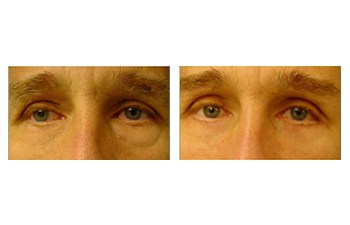
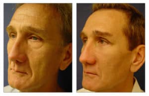
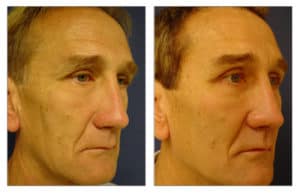
With meticulous surgical maneuvers and respect of vital structures of the lower eyelid anatomy, effective correction of the swollen lower eyelid can be achieved.
Please see this 52-year-old male following lower eyelid blepharoplasty and fat grafting to the cheeks.

